
Diffusion and Tractography
A comprehensive solution for diffusion and tractography analysis, providing detailed visualizations of neural pathways for enhanced clinical decision-making

The analysis of diffusion-weighted MRI data can be used to model water diffusion properties in the brain's white matter.
The nordicMEDiVA comprehensive solution for diffusion and tractography enables users to analyze MR diffusion data using both the DTI and Spherical Deconvolution (SD) models.
The improved SD model, based on the damped Richardson-Lucy spherical deconvolution algorithm by Dr. Flavio Dell’Acqua, allows for better tracking through areas of crossing fibers and gives a more complete representation of the anatomy. Watch the interview with Dr. Flavio Dell’Acqua to learn more!
DWI and Tracts
module
Advanced tractography is included
In addition to the calculation of diffusion tensor imaging (DTI) derived maps such as fractional anisotropy (FA) and mean diffusivity (MD), tractography can be performed to create 3D reconstructions of the major white matter pathways.
Fully customizable automatic pre-processing
In nordicMEDiVA, image processing pipelines are fully customizable and can be set up to trigger automatically. The Diffusion and Tractography pipeline includes:
3 model fitting algorithms: Diffusion Tensor Imaging (DTI), Spherical Deconvolution (SD), and Apparent Diffusion Coefficient (ADC).
Eddy current correction can be applied as a pre-processing step, which also includes motion correction.
Flexible export of Tractography results
Images can be sent as:
White pixels on grayscale — fused images for surgical planning.
Tractography DICOM — streamlines.

Flexible views
Choose from 10+ colors including the directional color option or add your custom colors. Adjust the streamlines displayed, and the length threshold for each tract shown, to clean up your data. With the "toggle visibility" action, easily select the tracts you wish to see.

Switch between different algorithms
Process maps using different model fitting algorithms and settings and review the tracts without recreating them each time.
ROIs approach for dissection
Visualize streamlines using regions of interest (ROIs) as dissection filters. AND (Waypoint), NOT (Exclusion), and END (Termination) logical operators are available.
Dynamic tracts visualization in 2D and 3D views
Dynamic visualization and adjustment of ROIs offer greater control and precision during analysis. This flexibility enhances the ability to trace neural pathways and modify regions on the fly.
See the difference!
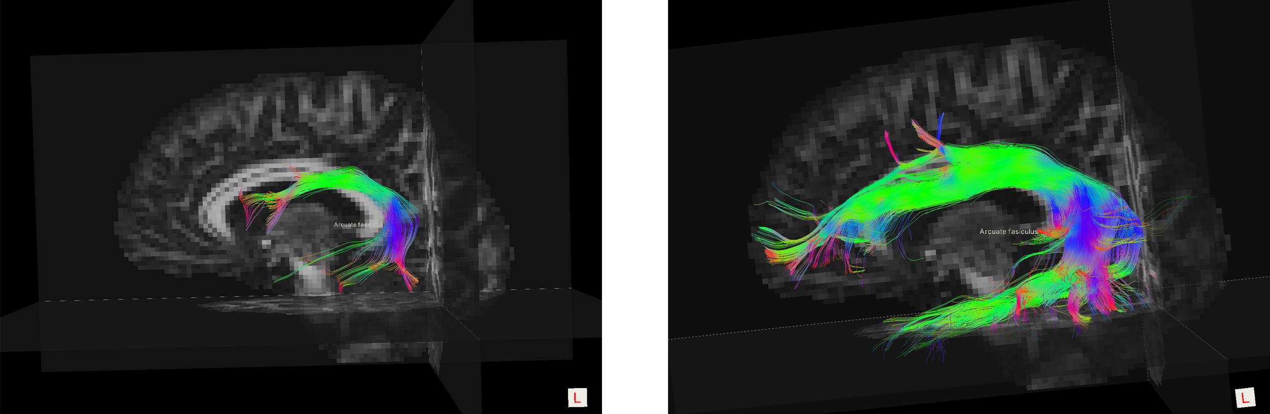
The images below show the corticospinal tract and the arcuate fasciculus from the same scan and patient.
Spherical deconvolution
algorithm
Classical diffusion tensor imaging
algorithm

We are committed to providing the highest quality on our product
-
Automation
The automatic routing feature in nordicMEDiVA is easy to set up and allows the system to automatically pre-process the raw data.
All this can be done within minutes from when the patient is scanned!
-
Cutting edge algorithms
The Spherical deconvolution algorithm implemented in nordicMEDiVA is a modified version of a damped Richardson-Lucy spherical deconvolution with deterministic Euler tractography to reduce isotropic background effects.
Push for innovation
Deliver high quality
Listen to our customers
Value safety
Focus on ease of use
Scientific references
Below you will find a list of scientific references, papers, and guidelines we have used in the development of the nordicMEDiVA task-based fMRI module. More references can be found on News & events | NordicNeuroLab
-
Abstract
Spherical deconvolution methods have been applied to diffusion MRI to improve diffusion tensor tractography results in brain regions with multiple fibre crossing. Recent developments, such as the introduction of non-negative constraints on the solution, allow a more accurate estimation of fibre orientations by reducing instability effects due to noise robustness. Standard convolution methods do not, however, adequately model the effects of partial volume from isotropic tissue, such as gray matter, or cerebrospinal fluid, which may degrade spherical deconvolution results. Here we use a newly developed spherical deconvolution algorithm based on an adaptive regularization (damped version of the Richardson-Lucy algorithm) to reduce isotropic partial volume effects. Results from both simulated and in vivo datasets show that, compared to a standard non-negative constrained algorithm, the damped Richardson-Lucy algorithm reduces spurious fibre orientations and preserves angular resolution of the main fibre orientations. These findings suggest that, in some brain regions, non-negative constraints alone may not be sufficient to reduce spurious fibre orientations. Considering both the speed of processing and the scan time required, this new method has the potential for better characterizing white matter anatomy and the integrity of pathological tissue.
-
Abstract
Diffusion tensor imaging (DTI) methods are widely used to reconstruct white matter trajectories and to quantify tissue changes using the average diffusion properties of each brain voxel. Spherical deconvolution (SD) methods have been developed to overcome the limitations of the diffusion tensor model in resolving crossing fibers and to improve tractography reconstructions. However, the use of SD methods to obtain quantitative indices of white matter integrity has not been extensively explored. In this study, we show that the hindrance modulated orientational anisotropy (HMOA) index, defined as the absolute amplitude of each lobe of the fiber orientation distribution, can be used as a compact measure to characterize the diffusion properties along each fiber orientation in white matter regions with complex organization. We demonstrate that the HMOA is highly sensitive to changes in fiber diffusivity (e.g., myelination processes or axonal loss) and to differences in the microstructural organization of white matter like axonal diameter and fiber dispersion. Using simulations to describe diffusivity changes observed in normal brain development and disorders, we observed that the HMOA is able to identify white matter changes that are not detectable with conventional DTI indices. We also show that the HMOA index can be used as an effective threshold for in vivo data to improve tractography reconstructions and to better map white matter complexity inside the brain. In conclusion, the HMOA represents a true tract‐specific and sensitive index and provides a compact characterization of white matter diffusion properties with potential for widespread application in normal and clinical populations. Hum Brain Mapp 34:2464–2483, 2013. © 2012 Wiley Periodicals, Inc.
-
Abstract
The diagonal and off-diagonal elements of the effective self-diffusion tensor, Deff, are related to the echo intensity in an NMR spin-echo experiment. This relationship is used to design experiments from which Deff is estimated. This estimate is validated using isotropic and anisotropic media, i.e., water and skeletal muscle. It is shown that significant errors are made in diffusion NMR spectroscopy and imaging of anisotropic skeletal muscle when off-diagonal elements of Deff are ignored, most notably the loss of information needed to determine fiber orientation. Estimation of Deff provides the theoretical basis for a new MRI modality, diffusion tensor imaging, which provides information about tissue microstructure and its physiologic state not contained in scalar quantities such as T1, T2, proton density, or the scalar apparent diffusion constant.
-
Abstract
Purpose: Linear least squares estimators are widely used in diffusion MRI for the estimation of diffusion parameters. Although adding proper weights is necessary to increase the precision of these linear estimators, there is no consensus on how to practically define them. In this study, the impact of the commonly used weighting strategies on the accuracy and precision of linear diffusion parameter estimators is evaluated and compared with the nonlinear least squares estimation approach.
Methods:Simulation and real data experiments were done to study the performance of the weighted linear least squares estimators with weights defined by (a) the squares of the respective noisy diffusion-weighted signals; and (b) the squares of the predicted signals, which are reconstructed from a previous estimate of the diffusion model parameters.
Results: The negative effect of weighting strategy (a) on the accuracy of the estimator was surprisingly high. Multi-step weighting strategies yield better performance and, in some cases, even outperformed the nonlinear least squares estimator.
Conclusion: If proper weighting strategies are applied, the weighted linear least squares approach shows high performance characteristics in terms of accuracy/precision and may even be preferred over nonlinear estimation methods.
-
Abstract
Fiber tract trajectories in coherently organized brain white matter pathways were computed from in vivo diffusion tensor magnetic resonance imaging (DT-MRI) data. First, a continuous diffusion tensor field is constructed from this discrete, noisy, measured DT-MRI data. Then a Frenet equation, describing the evolution of a fiber tract, was solved. This approach was validated using synthesized, noisy DT-MRI data. Corpus callosum and pyramidal tract trajectories were constructed and found to be consistent with known anatomy. The method's reliability, however, degrades where the distribution of fiber tract directions is nonuniform. Moreover, background noise in diffusion-weighted MRIs can cause a computed trajectory to hop from tract to tract. Still, this method can provide quantitative information with which to visualize and study connectivity and continuity of neural pathways in the central and peripheral nervous systems in vivo, and holds promise for elucidating architectural features in other fibrous tissues and ordered media. Magn Reson Med 44:625–632, 2000. Published 2000 Wiley-Liss, Inc.
https://onlinelibrary.wiley.com/doi/10.1002/1522-2594(200010)44:4%3C625::AID-MRM17%3E3.0.CO;2-O
-
Abstract
The relationship between brain structure and complex behavior is governed by large-scale neurocognitive networks. The availability of a noninvasive technique that can visualize the neuronal projections connecting the functional centers should therefore provide new keys to the understanding of brain function. By using high-resolution three-dimensional diffusion magnetic resonance imaging and a newly designed tracking approach, we show that neuronal pathways in the rat brain can be probed in situ. The results are validated through comparison with known anatomical locations of such fibers. Ann Neurol 1999;45:265–269
https://onlinelibrary.wiley.com/doi/10.1002/1531-8249(199902)45:2%3C265::AID-ANA21%3E3.0.CO;2-3